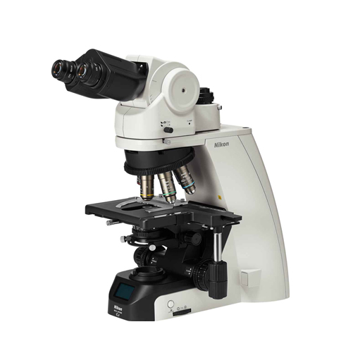
Inverted microscope system
Leading advanced imaging platform
ECLIPSE Ti2 offers an unparalleled 25mm field of view (FOV), completely changing your perception. With this incredible field of view, Ti2 can maximize the sensor area of large target CMOS cameras without compromising elsewhere and significantly improve data throughput. Ti2's highly stable and drift free platform is designed to meet the needs of super-resolution imaging, while its unique hardware triggering capabilities can even enhance the most challenging high-speed imaging applications. In addition, Ti2's unique intelligent features guide users through the imaging workflow by collecting data from internal sensors, thereby eliminating the possibility of user errors. In addition, the status of each sensor is automatically recorded during the acquisition process, providing quality control for imaging experiments and enhancing data reproducibility.
Combined with Nikon's powerful acquisition and analysis software NIS Elements, Ti2 has become a new innovation in the imaging field.
A pioneering broad perspective
As research trends shift towards large-scale system level methods, the demand for faster data collection and higher throughput capabilities continues to increase. The development of sensors for large target area cameras and the improvement of PC data processing capabilities have promoted this research trend. Ti2 has an unprecedented 25mm field of view, providing a higher level of scalability that allows researchers to truly maximize the practicality of large target area detectors and provide future oriented capabilities for its core imaging platform while camera technology continues to rapidly develop.
Provide bright illumination in a wide field of view
High power LEDs can provide bright illumination within the wide field of view of Ti2, ensuring clear and consistent results from demanding applications such as high magnification DIC. The design incorporating compound eye lenses provides uniform illumination from the center to the edges for seamless image stitching in quantitative high-speed imaging and magnification applications.
A compact epifluorescence illumination device designed specifically for large field imaging, equipped with a quartz compound eye lens that provides high transmittance over a wide spectrum including ultraviolet light. A large-diameter fluorescent filter with a hard coating can provide a large field of view image with high signal-to-noise ratio.
Large diameter observation optical system
The diameter of the observation light path has been expanded to achieve a field of view of 25 at the imaging port. The resulting large field of view can capture approximately twice the area of traditional optical devices, allowing users to achieve maximum performance from large sensors such as CMOS detectors.
Objective lens for large field imaging
Objective lenses with excellent image flatness ensure high-quality images from center to edge. The maximum potential of utilizing OFN25 objective lens can significantly accelerate data collection.
Camera for large capacity data acquisition
The Nikon FX format F-interface cameras Digital Sight 50M and Digital Sight 10 are equipped with CMOS image sensors originally developed for D-SLR professional cameras, optimized for research purposes. It can achieve high-speed and high-sensitivity live cell imaging and the most effective utilization of Ti2 large field of view.
Unparalleled Nikon optical system
Nikon's high-precision CFI60 infinity optical device is designed for various complex observation methods and has been highly praised by researchers for its excellent optical performance and robust reliability.
Cut toe difference
Nikon's unique apodization objective and optional amplitude filter can significantly improve contrast and reduce halo artifacts, providing detailed high-definition images
External differences
The electric external phase difference system enables users to combine phase difference with epifluorescence imaging without affecting fluorescence transmission by bypassing the need for phase difference objectives. For example, liquid immersion objectives with very high numerical apertures can be used for phase contrast imaging. By using this external phase difference system, users can easily combine phase difference with other imaging modes, including weak fluorescence imaging for applications such as TIRF and laser tweezers
DIC (Differential Interference Difference)
Nikon's highly acclaimed DIC optical components provide uniform, clear, and detailed images across the entire magnification range, with high resolution and contrast. DIC prisms are individually customized for each objective lens to provide the highest quality DIC images for each sample.
NAMC (Nikon Advanced Modulation Contrast)
This is a plastic compatible high contrast imaging technique suitable for unstained transparent samples such as oocytes. NAMC provides pseudo 3D images with a shadow cast appearance. Users can easily adjust the direction of contrast for each sample.
Automatic correction loop
The thickness of the sample, the thickness of the cover glass, the refractive index distribution in the sample, and temperature changes can cause spherical aberration and image distortion. The highest quality objective lenses are usually equipped with correction rings to compensate for these variations. And precise adjustment of the correction ring is crucial for achieving high-resolution and high contrast images. This new type of automatic correction ring adopts harmonic drive and automatic correction algorithm, allowing users to easily achieve precise correction ring adjustment to achieve optimal performance of the objective lens every time.
Epifluorescence
The Lambda series objective lens adopts Nikon's proprietary nanocrystalline coating technology, which is very suitable for high, low signal, multi-channel fluorescence imaging that requires high transmission and aberration correction over a wide wavelength range. The Lambda series objective lenses have demonstrated their ability in weak signal observation (such as single-molecule imaging) and even luminescence based applications, by combining new fluorescence filtering cubes that provide improved fluorescence detection and stray light countermeasures (such as noise terminators).
Volume Contrast
Volume Contrast utilizes a series of unlabeled bright field images captured at different Z-axis depths to reconstruct phase distribution images.
Volume Contrast images facilitate cell recognition and provide convenience for automatic counting and area analysis. Due to the use of bright field imaging, VC can perform real-time and non-destructive analysis of cells, making it suitable for various applications. (Only applicable to TI2-E).
Volume Contrast feature
Accurately identify cells from unlabeled samples for automatic cell counting and area measurement.
Eliminating the impact of the half moon effect on cell identification
Due to the half moon effect, the phase difference image is adversely affected at the edge of the culture well. VC avoids this impact, allowing cells at the edge of the culture well to be clearly identified, thereby increasing cell counting accuracy and improving statistical data.
Perfect focus
Even the slightest changes in temperature and vibration in the imaging environment can greatly affect focusing stability. Ti2 uses static and dynamic measurements to eliminate focal drift, enabling faithful visualization of the nanoscale and microscopic worlds in long-term experiments.
Mechanical redesign of Ti2-E to achieve ultra-high stability (Ti2-E)
In order to improve focusing stability, both the Z-axis drive and PFS autofocus mechanism have undergone a comprehensive redesign.
The new Z-axis focusing mechanism is smaller and located near the objective lens turntable, which can minimize vibration to the greatest extent possible. Even with an extended (double-layer optical path) configuration, it still remains near the objective turntable, ensuring stability for all applications.
The detector part of the Perfect Focus System (PFS) has been removed from the objective disc to reduce the mechanical load on the objective disc. This new design can also minimize heat transfer, thereby helping to achieve a more stable imaging environment. As a result, the power consumption of the Z-axis drive motor has also been reduced. These mechanical redesigns combined form an ultra stable imaging platform that is highly suitable for single-molecule imaging and super-resolution applications.
Real time focus correction using PFS: Simple and perfect Ti2-E
The Perfect Focus System (PFS) can automatically correct focus drift caused by temperature changes and mechanical vibrations, which may be caused by various factors, including adding reagents to the sample and multi position imaging.
PFS maintains focus by real-time detection and tracking of the position of the cover glass surface. The unique optical offset technology allows users to easily maintain the focus at the desired position off the surface of the cover glass. PFS automatically maintains continuous focus through a built-in linear encoder and high-speed feedback mechanism, providing highly reliable images even in long-term complex imaging tasks.
PFS is compatible with a wide range of applications, from routine experiments involving plastic culture dishes to single-molecule imaging and multiphoton imaging. It is also compatible with various wavelengths from ultraviolet to infrared, which means it can be used for multiphoton and optical tweezers applications.
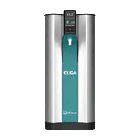
Objective automatic water replenishment device Ti2-E
The use of a new objective automatic water replenishment device can improve the long-term imaging performance of PFS and water mirrors. The automatic water replenishment device for the objective lens automatically applies an appropriate amount of pure water to the tip of the objective lens to prevent it from becoming dry and overflowing during the experiment. It is compatible with all types of water mirrors, helping to provide high-resolution, high contrast, and aberration corrected delayed images stably over long periods of time.
Compatible objectives
CFI Apochromat LWD Lambda S 20XC WI
CFI Apochromat Lambda S 40XC WI
CFI Apochromat LWD Lambda S 40XC WI
CFI Plan Apochromat VC 60XC WI
CFI Plan Apochromat IR 60XC WI
CFI SR Plan Apochromat IR 60XC WI
CFI SR Plan Apochromat IR 60XAC WI
Auxiliary guide
Users no longer need to memorize complex microscope alignment and operation procedures. Ti2 integrates data from sensors to guide you through these steps, reduce user errors, and enable researchers to focus on their data.
Continuous display of microscope status Ti2-E/A
A series of built-in sensors can detect and transmit status information of various components in the microscope. When using a computer to obtain images, all status information is recorded in metadata, so you can easily call collection conditions and/or check for configuration errors.
In addition, the built-in camera allows users to view the back focal plane, making it easy to calibrate the phase difference ring and DIC extinction cross. It also provides laser safe alignment methods for applications such as TIRF.
The microscope status can be viewed on the tablet or displayed through the indicator lights in front of the microscope, making it easy to confirm the device status in the dark room.
Operation Procedure Wizard Ti2-E/A
The auxiliary guide function of Ti2 provides interactive step-by-step guidance for microscope operation. You can view the auxiliary guide on your tablet or PC and integrate real-time data from built-in sensors and cameras. The auxiliary guide aims to assist users in completing the calibration procedure for experimental setup and troubleshooting.
Automatically detect errors Ti2-E/A
The inspection mode allows users to easily confirm whether all correct microscope components are suitable for their chosen observation method on a tablet or PC. When the required observation method is not implemented, this feature eliminates the time and effort typically required for troubleshooting. This feature is particularly advantageous when multiple users are involved, as each user may make unexpected changes to the microscope settings. Custom inspection programs can also be pre programmed.
Intuitive operation
Ti2 has undergone a comprehensive redesign, from the overall design to the selection and placement of each button and switch, bringing users the ultimate experience. Even in the dark, control is easy to use and most imaging experiments are conducted in the dark. Ti2 provides an intuitive and easy-to-use user interface, allowing researchers to focus on data rather than microscope control.
Carefully designed microscope control layout Ti2-E Ti2-A
The positions of all buttons and switches are based on the type of lighting they control. The button for controlling transmission observation is located on the left side of the microscope, and the button for controlling fluorescence observation is located on the right side. The buttons for controlling common operations are located on the front panel. The use of this partition provides an easy to remember layout, which is an ideal feature when operating a microscope in a dark room.
① Reciprocating switching (Ti2-E)
The design incorporates reciprocating switching for controlling devices such as the fluorescent filter turntable and objective lens turntable. These types of switches mimic the feeling of manually rotating these devices to achieve intuitive control. Additional functions can be integrated into these reciprocating switches, allowing a single switch to operate multiple related devices. For example, the reciprocating switching used for the fluorescent filter turntable not only rotates the turntable, but also opens and closes the fluorescent shutter when the user presses the switch. These switches can also be programmed to operate the emission filter dial and external phase difference unit.
② Programming Function Button (Ti2-E/A)
The conveniently located function buttons support custom user interfaces. Users can choose from over 100 functions, including controlling electric devices such as shutters, and even outputting signals to external devices through I/O ports for triggering acquisition. By storing the settings of each electric device, the mode function that can instantly change the observation method can also be assigned to these buttons.
③ Focus knob (Ti2-E)
The focus acceleration button and PFS engagement button are set near the focus knob. Due to their different shapes, these two buttons are easily recognizable by touch. The focusing speed is automatically adjusted according to the use of the objective lens, achieving pressure free operation by maintaining the ideal focusing speed.
Use joystick and tablet for intuitive control
The Ti2 joystick not only controls the movement of the stage, but also controls most of the electric functions of the microscope, including PFS activity. It can display XYZ coordinates and the status of microscope components, providing an effective means for users to remotely control the microscope. The electric function of Ti2 can also be controlled through a tablet computer and connected to the microscope via wireless LAN, providing a universal graphical interface for microscope control.
Ti2 Control: a dedicated application for smartphones and tablets
It can set and control the settings, status display, and operation guidance of Ti2-E and Ti2-A.
download
TI2 control can be accessed from the App Store ® And Google Play ™ Free download.
Download it to an iPhone like device ®、iPad ® Or Android ™ On the device. For information on compatible devices, please refer to the "Operating Conditions" section below.
Operating conditions
IOS devices: devices equipped with iOS 10.3 or higher (iPhone 7 or higher, iPad Air 2 or higher)
Android ™ Device: Equipped with Android 5.1 or higher version
Apple ®, App Store ®, Apple logo, iPhone ® And iPad ® It is a registered trademark of Apple Inc. in the United States and other countries/regions.
The iOS trademark is used under license in the United States and other countries/regions.
The iPhone trademark is used under license from Aiphone Co. Ltd.
Android ™ And Google Play ™ It is a registered trademark of Google Inc.

Phone: 0512-69321899
QQ:3463428168
Mobile phone: 18913550679
Address: A611 Chenlei Technology Park, No.1 Qunxing 1st Road, Suzhou Industrial Park, Jiangsu Province
Get company updates
 Click to learn